Intravitreal Injection Guide: A Primer for Ophthalmology
Over the last few years, the use of Intraocular Injections or Intravitreal Injections has become the routine standard-of-care for various retinal procedures in ophthalmology. An intravitreal injection is a method to inject a higher level of the medication directly into the vitreous chamber, near the retina at the back of the eye, which is filled with a jelly-like fluid known as the vitreous humor gel. The injected medicine can help to protect the vision by treating various eye problems. However, the use of a drape and speculum for injecting the medicine into the eye can be unpleasant for many patients. Studies have indicated that the use of drape results in feelings of anxiety, claustrophobia, and a high level of stress among the patients along with pain during the needle insertion. Also, the touching of the needle tip to the eyelashes and eyelid can cause inadequate sterility and contamination resulting in infections like endophthalmitis.
 To overcome these issues, Malosa Intravitreal Injection Guide (IVT) has been developed that features an in-built lash guard which eliminates the need for a surgical drape and speculum during the IVT injection procedure. The use of the Intravitreal Injection Guide is expected to deliver safe, faster, and accurate injections.
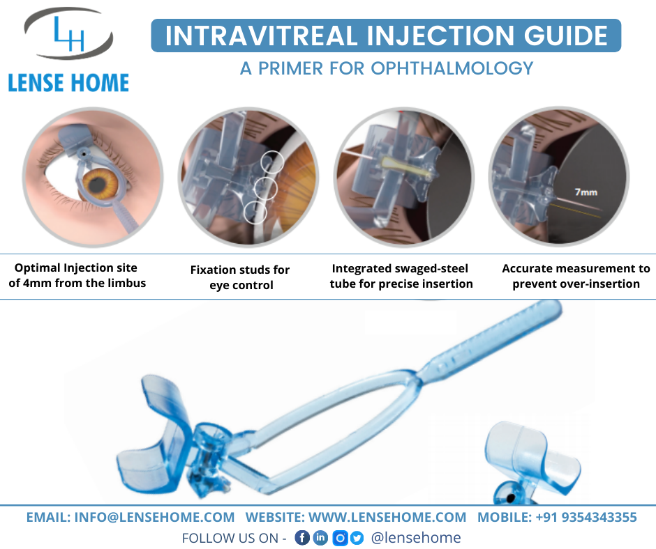
The Malosa Intravitreal Injection Guide is a single-use, polycarbonate, and sterile instrument that comprise a triangular footplate, a handle, a lash guard, and a needle chamber. The fixation studs and an Apex indicator present in the footplate are to ensure globe stabilization and accuracy in limbal placement. The handle comes in a wishbone shape that ensures enough visualization of the procedure and the cylindrical needle chamber facilitates controlled and precise entry of the needle at a fixed depth. The chamber is designed in such a manner that it can be used with a 30-gauge needle for providing added comfort to the patient and minimizing the pain and discomfort during needle insertion.
The fixation studs present on the footplate of the guide helps in controlling the patient’s eye movements occurring due to uncomfortable sensation or pre-procedure nerves related to the use of a lid speculum. The eyes can get stable with the instrument once the patient looks in the required direction. With this, unnecessary risks like retinal damage and corneal abrasions to the patient can be prevented.
Improving Technique’s Safety Profile with IVT Guide: The use of the Intravitreal Injection Guide can help in the standardization of the procedure that can result in the reduction of complications like traumatic lens injury. In the standard procedure, the tip of the needle requires to be pointed towards the center of the globe while the needle is inserted through the sclera in a perpendicular direction for preventing any injury to the posterior. The injection site is then marked using sterile ophthalmic calipers. To overcome these challenges, the IVT Guide is exclusively designed to have an arrow on the apex of the plate base for accurate positioning at the limbus. The cylindrical chamber ensures that there is no over-insertion of the needle and allows only 7-mm insertion of a 13-mm/30-Gauge Needle into the eye. The guide ensures that the injection is delivered at an accurate perpendicular angle to the sclera and a correct distance of 4mm from the limbus.
The increasing demand for intravitreal injections increases the need for a device like IVT Guide that can reduce the workload and make the procedure more efficient. Considering such practical implications of the device and the level of patient satisfaction, the use of the IVT Guide foresees a bright future in ophthalmology.


Leave a Reply